Welcome to the Smolikove lab at the University of Iowa
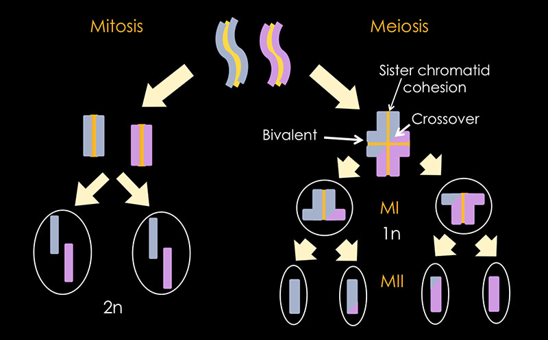
We are interested in understanding the crucial steps leading to faithful chromosome segregation during meiosis. Meiosis is a specialized cell division resulting in the production of gametes (e.g. eggs and sperm). This division concludes in the reduction of the number of chromosomes by half.
Upon fertilization, the number of chromosomes is restored so that it is identical to the one found in the parental cells. The accurate reconstitution of chromosome numbers is crucial for the development of a healthy embryo. Alterations in chromosome numbers are the leading known causes for serious birth defects including Down syndrome as well as miscarriages and stillbirths. Therefore, the accurate separation of homologous chromosomes during meiosis is key event for successful sexual reproduction and is of tremendous importance for human health.
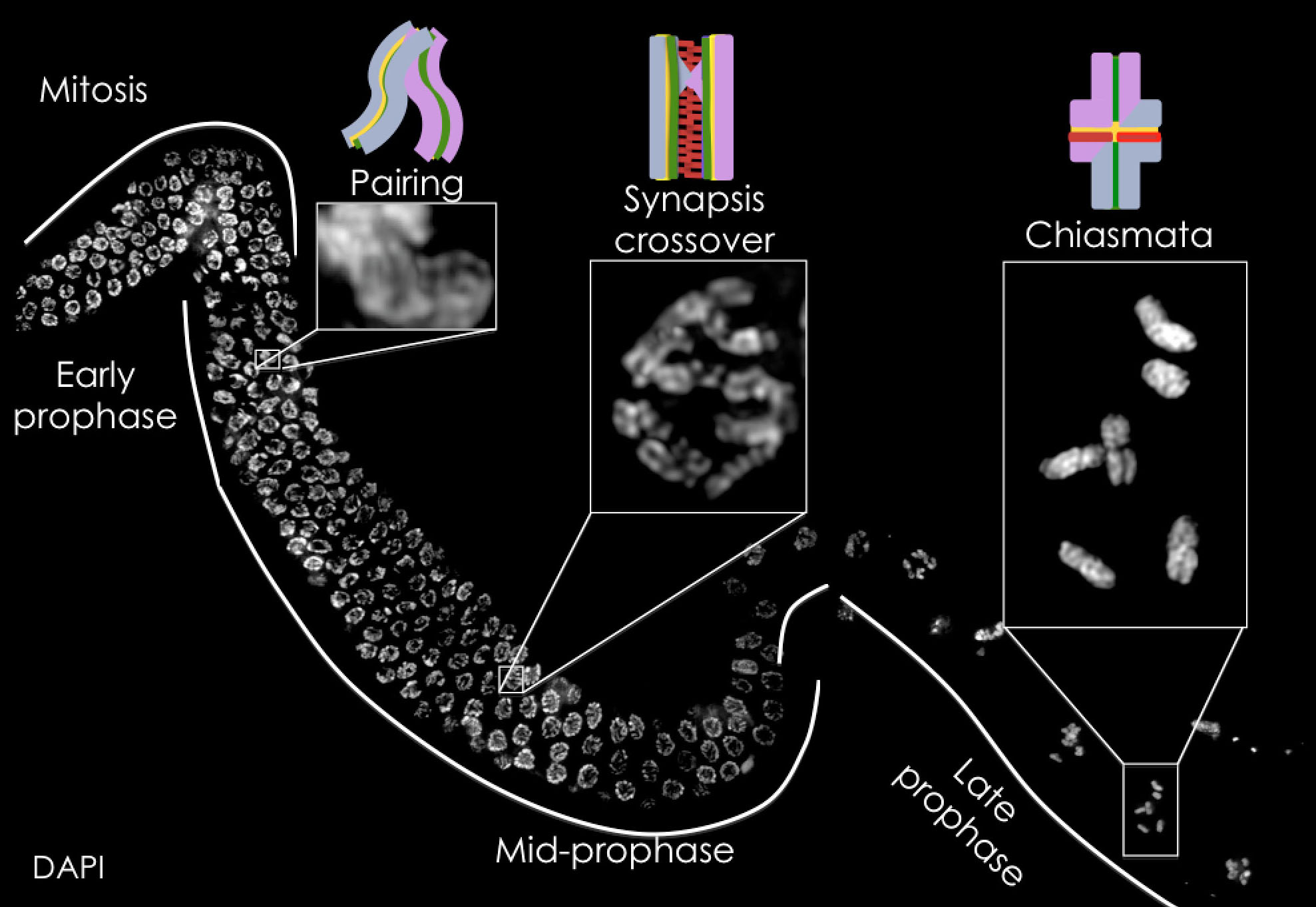
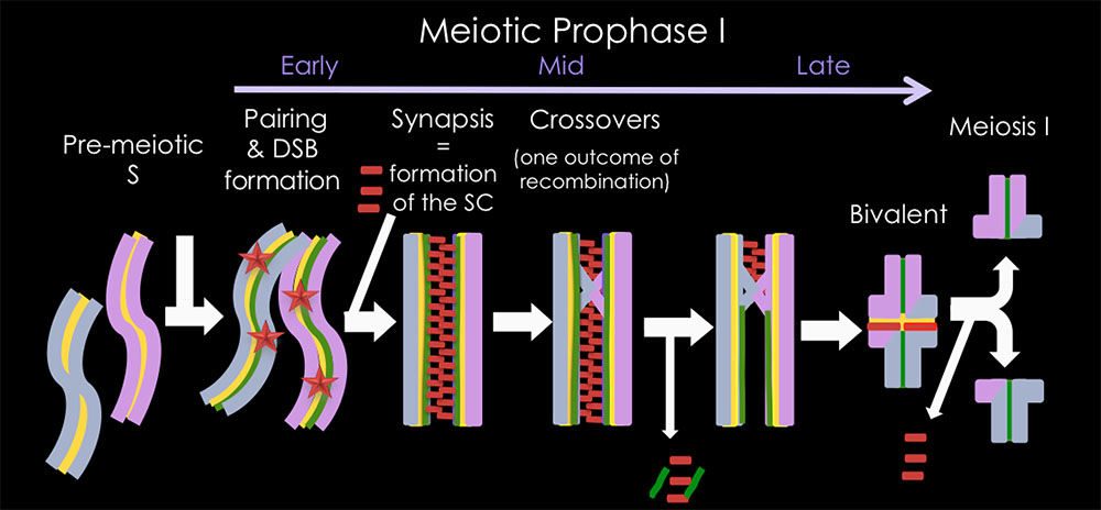
During meiotic prophase I, the two homologous chromosomes (one each of paternal and of maternal origin) separate (see figure above). Formation of a physical link between homologous chromosomes prior to their segregation is required for this accurate separation. This link is mediated via crossovers, the exchange of genetic material between the homologous chromosomes.
To ensure the production of at least one obligatory crossover, the DNA of the meiotic prophase I germ cell (blue and purple in the figure below) is broken by the protein SPO-11 (stars in the figure below). These meiotic double stand breaks (DSBs) are repaired in the presence of the synaptonemal complex (SC, red bars in the figure below) to form crossovers. The SC is formed between each pair of homologous chromosomes. In the absence of a functional SC or the repair of DSBs to form crossovers chromosomes missegregate in cells that proceed to the meiotic division.
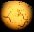
For our studies of meiosis, we use Caenorhabditis elegans as a model system (figure bel0w). C. elegans is a microscopic worm, which is a well-established model organism amenable for a variety of research approaches including genetic manipulation, cell biology and biochemistry.
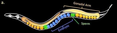
We can analyze chromosomes in the live organisms as well as in dissected gonads. Meiotic prophase nuclei are the majority of nuclei composing the germline of this microscopic worm (see figure below). These nuclei are arranged in temporal-spatial pattern; therefore, all early meiotic events can be analyzed simultaneously in a single gonad. We use genetic and cell biology techniques involving high-resolution microscopy to study how chromosomes behave in these early meiotic events.